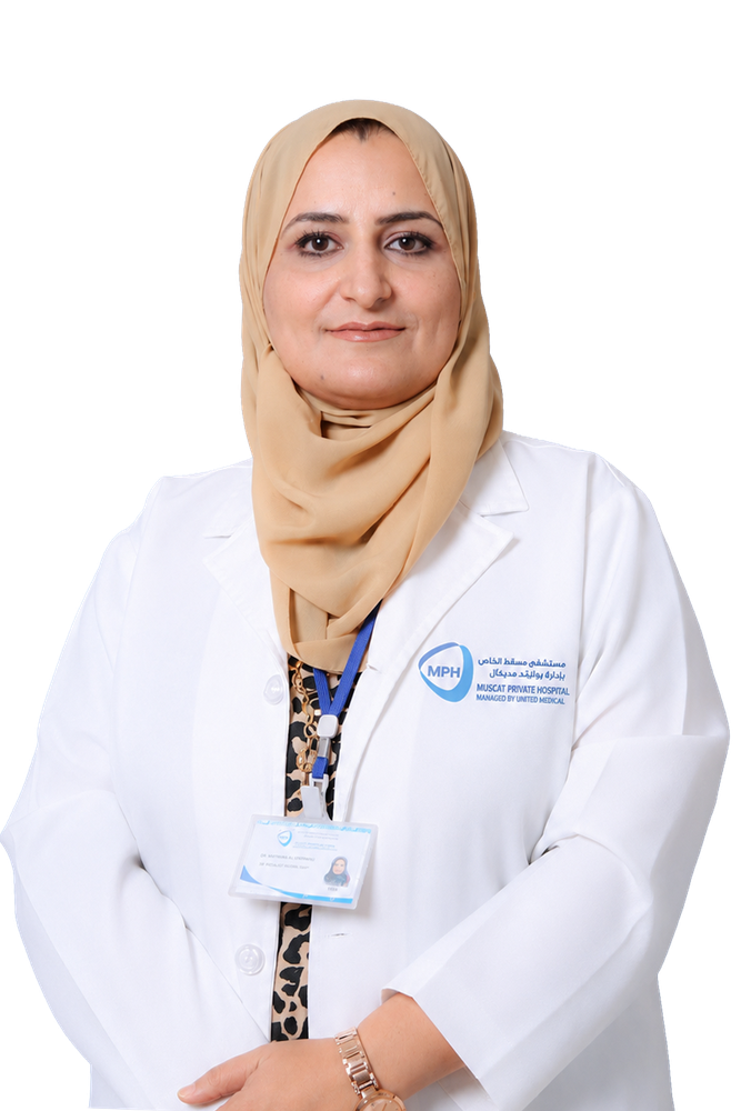
About Dr. Shaymaa
Dr. Shaymaa is a Specialist Diagnostic Radiologist with 20 years of experience in diagnostic imaging and image-guided procedures. Her reporting practice covers ultrasound, mammography, bone density (DXA), general radiography, fluoroscopy and computed tomography.
She qualified in medicine at the University of Baghdad, and went on to complete a Diploma in Diagnostic Radiology there in 2012. She holds the European Diploma in Radiology (EDiR), awarded by the European Board of Radiology in 2021, and has passed Part 1 of the Fellowship examination of the Royal College of Radiologists.
A substantial part of Dr. Shaymaa's work is in women's imaging. She reports first-trimester screening including early pregnancy assessment and nuchal translucency, detailed second-trimester anomaly scans, third-trimester growth and biophysical profile studies, and fetal Doppler surveillance, together with 3D and 4D obstetric ultrasound. Her gynaecological work includes the assessment of adnexal masses, endometriosis mapping, uterine anomalies, follicular tracking, and hysterosalpingography for infertility and tubal patency.
Alongside this she reports breast imaging across digital mammography, tomosynthesis and breast ultrasound; paediatric ultrasound including neonatal cranial studies and acute paediatric presentations; adult multi-system ultrasound; vascular and Doppler studies; and whole-body CT in emergency and elective settings. She also performs ultrasound-guided interventions including core biopsy, fine-needle aspiration and wire localisation.
Dr. Shaymaa has a particular interest in structured, standardised reporting using systems such as BI-RADS, TI-RADS and O-RADS, in quality assurance for mammography screening, in radiation safety, and in workflow design for hospital imaging units. She works closely with referring clinicians across specialties to make sure imaging findings translate into clear next steps for the patient.
Patient-Care Philosophy
“I am committed to delivering the highest standard of diagnostic accuracy while prioritising patient safety, comfort and radiation optimisation (ALARA). I believe radiology is the cornerstone of effective medicine, requiring close collaboration with referring physicians to ensure precise, individualised patient pathways. My philosophy centres on treating the patient, not just the image — combining diagnostic care with compassionate communication, so that patients feel heard and respected during anxious moments.”
Areas of Expertise
Women's Imaging
Obstetric
First-trimester screening, including early pregnancy assessment and nuchal translucency
Second-trimester detailed anomaly scanning
Third-trimester growth assessment and biophysical profile
Fetal and obstetric Doppler studies
High-risk pregnancy surveillance, including fetal growth restriction, placental disorders and multiple pregnancy
3D and 4D obstetric ultrasound
Women's Imaging
Gynaecological
Assessment of adnexal masses
Endometriosis mapping
Uterine anomalies
Transabdominal and transvaginal pelvic ultrasound, including follicular tracking
Hysterosalpingography (HSG) for infertility and tubal patency assessment
Breast Imaging
Digital mammography and tomosynthesis
Breast ultrasound
Structured reporting using the BI-RADS system
Paediatric Imaging
Neonatal cranial ultrasound
Infant and paediatric abdominal ultrasound
Pyloric stenosis and intussusception
Acute paediatric presentations, including appendicitis, testicular and ovarian torsion, and mesenteric lymphadenitis
Adult General and Musculoskeletal Imaging
Abdominal and pelvic ultrasound
Small parts ultrasound, including thyroid and soft tissue
Musculoskeletal ultrasound
Skeletal, chest and acute abdominal radiography
Bone density assessment (DXA)
Vascular and Doppler Imaging
Carotid and vertebral Doppler
Renal artery Doppler
Portal and hepatic vein Doppler
Aortic assessment
Peripheral arterial and venous Doppler of the limbs
Emergency and Acute Imaging
Whole-body and high-resolution CT in the emergency setting
CT pulmonary angiography
Point-of-care ultrasound (POCUS) for critical care and emergency assessment
Procedures & Services
Ultrasound
Breast ultrasound
Obstetric ultrasound
First, second and third trimester, biophysical profile, obstetric Doppler, and 3D/4D fetal studies
Gynaecological ultrasound
Transabdominal and transvaginal pelvic scanning and follicular studies
Neck ultrasound
Thyroid, parathyroid, salivary glands and lymph node mapping
Adult abdominal ultrasound
Liver, gall bladder and bile ducts, spleen, pancreas, gastrointestinal tract, adrenals, kidneys and urinary bladder
Paediatric ultrasound
Neonatal brain, adrenals, uterus and ovaries, liver, gall bladder and bile ducts, spleen, pancreas, gastrointestinal tract, kidneys, urinary bladder and scrotum
Prostate ultrasound
Soft tissue ultrasound, including inguinal, umbilical and gluteal regions
Musculoskeletal ultrasound
Vascular and Doppler studies
Portal vein, hepatic vessels, aorta, carotid and vertebral arteries, renal arteries, and limb arterial and venous studies
Image-Guided Procedures
Ultrasound-guided fine-needle aspiration cytology (FNAC)
Ultrasound-guided core biopsy
Image-guided wire localisation
Breast Imaging and Bone Density
Mammography
Performance and reporting
Digital breast tomosynthesis
Bone density assessment (DXA)
Reporting
X-Ray and Fluoroscopy
Conventional diagnostic radiography
Hysterosalpingography (HSG)
Micturating cystourethrography (MCU)
Urethrography
Intravenous pyelography (IVP)
Other fluoroscopic studies
Computed Tomography (CT)
CT of the head, neck and brain
CT of the spine
CT of the chest, abdomen and pelvis
Musculoskeletal CT
CT pulmonary angiography
Whole-body CT
Education & Training
European Diploma in Radiology (EDiR)
European Board of Radiology
2021FRCR Part 1
Royal College of Radiologists, UK
Diploma in Diagnostic Radiology (DMRD)
College of Medicine, University of Baghdad, Iraq
2012MBChB (Bachelor of Medicine, Bachelor of Surgery)
College of Medicine, University of Baghdad, Iraq
2003
Frequently Asked Questions
Book Your Consultation with
Dr. Shaymaa in Muscat
Book your consultation with Dr. Shaymaa in Muscat, Oman. Our team is here to help you arrange the right appointment.
Prefer to speak with us? Call Dr. Shaymaa and our team will assist with appointment availability and insurance enquiries.